


 English
 عربى
عربى
 Español


×
Password
Get password
Enter password to download relevant content.
Submit
 +86-15267462807
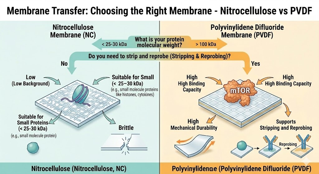
Direct answer: For most Western blots, PVDF is the safer default — it has higher protein binding capacity (170–200 µg/cm² vs 80–100 µg/cm²), better mechanical durability, and supports stripping and reprobing. But nitrocellulose is not inferior — it has lower background, no methanol activation step, and is better for small proteins (< 25–30 kDa). The right choice depends on your target protein’s size and abundance, your detection method, and whether you need to reprobe. Neither membrane is universally “better.”
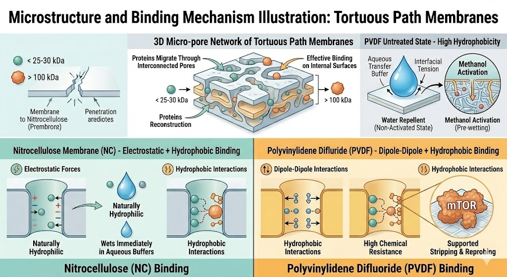
Both nitrocellulose and PVDF are tortuous path membranes — proteins migrate through a three-dimensional network of interconnected pores and bind to the internal surface area, not just the outer face. This structure gives both membranes far higher effective binding surface than their flat dimensions suggest.
The binding mechanism differs:
This difference in wetting behavior is the most common source of PVDF transfer failure in the lab. A PVDF membrane that dries out mid-experiment must be re-wetted before continuing.
| Parameter | Nitrocellulose | PVDF |
|---|---|---|
| Protein binding capacity | 80–100 µg/cm² | 170–200 µg/cm² |
| Binding mechanism | Hydrophobic + electrostatic | Hydrophobic + dipole–dipole |
| Methanol pre-wetting required | No | Yes |
| Mechanical durability | Fragile, tears easily | Tough, chemically resistant |
| Background noise | Low | Moderate (higher with fluorescence) |
| Sensitivity (low-abundance proteins) | Moderate | High |
| Best MW range | Low MW (< 25–30 kDa) | High MW (> 100 kDa) |
| Stripping and reprobing | Difficult — signal loss | Excellent |
| Fluorescence detection | Not recommended (high autofluorescence) | Yes — use low-fluorescence PVDF |
| Mass spectrometry (MS) downstream | No | Yes |
| Protein sequencing (Edman degradation) | No | Yes |
| Nucleic acid blotting (DNA/RNA) | Yes | No |
| Relative cost | Lower | Higher |
The most commonly misunderstood aspect of membrane selection is the relationship between molecular weight and membrane choice.
The conventional advice — “use PVDF for sensitive detection, nitrocellulose for routine work” — misses a critical nuance. A 2021 systematic study published in Scientific Reports compared binding ability of both membranes across proteins of low, medium, and high molecular weight. The findings were:
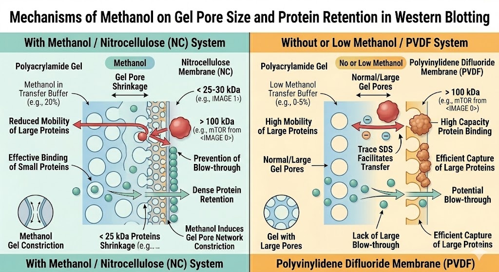
The reason relates to transfer buffer composition. Nitrocellulose transfer protocols typically include methanol in the transfer buffer. Methanol reduces gel pore size during electrotransfer, which prevents small proteins from running back through the gel — improving retention of small proteins on the membrane. However, this same methanol reduces the mobility of large proteins out of the gel, impairing their transfer efficiency for high MW targets.
PVDF does not require methanol in the transfer buffer. Without methanol, large proteins transfer more efficiently — which is why PVDF consistently outperforms nitrocellulose for proteins above 100 kDa.
Practical MW selection guide:
| Target protein MW | Recommended membrane | Reason |
|---|---|---|
| < 15 kDa (small peptides) | Nitrocellulose (0.2 µm) | Better small-protein retention; methanol in buffer helps |
| 15–30 kDa | Nitrocellulose or PVDF | Either acceptable; NC slightly preferred |
| 30–100 kDa | PVDF | Higher binding capacity, reliable detection |
| > 100 kDa | PVDF (methanol-free transfer buffer) | NC + methanol impairs large protein transfer |
| Multiple targets spanning wide MW range | PVDF | More consistent across the full range |
Both membranes are available in three standard pore sizes. Pore size is a separate decision from membrane material — choose both independently.
| Pore Size | Best For | Notes |
|---|---|---|
| 0.1 µm | Proteins < 10 kDa, very small peptides | Highest retention, highest background risk |
| 0.2 µm | Proteins < 20 kDa; low-loading quantitative work | Good balance for small proteins |
| 0.45 µm | Proteins > 20 kDa; standard applications | Default for most Western blots |
Rule: When your target protein is small (< 15 kDa) or your loading amount is low and quantification is critical, always use 0.2 µm rather than 0.45 µm — regardless of membrane material. The smaller pore size reduces protein run-through during transfer.
Membrane choice must align with your detection strategy.
Both membranes are fully compatible. This is the most common detection method and the least discriminating — either membrane works. If all other factors are equal and you’re using ECL, choose based on protein MW and reprobing needs.
Use low-fluorescence PVDF. Standard nitrocellulose has high autofluorescence that bleeds into fluorescence detection channels — it produces elevated background that obscures weak signals and makes two-color multiplexing unreliable. Standard PVDF also has moderate autofluorescence. For fluorescence-based Western blot (e.g., LI-COR Odyssey systems), specify low-fluorescence PVDF explicitly — it is a distinct product category, not just standard PVDF.
Both membranes are compatible. Nitrocellulose tends to give lower background with colorimetric substrates due to better blocking characteristics.
Both compatible. Nitrocellulose is the historical standard for radioactive detection and slightly preferred for this application.
If your experiment requires probing the same membrane with more than one primary antibody — whether sequentially for different targets or after stripping to reprobe with a loading control — membrane durability becomes critical.
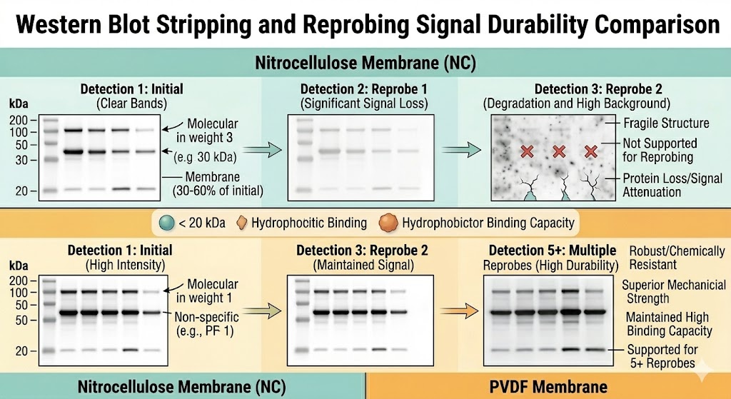
Standard nitrocellulose is fragile. Stripping protocols involving high-temperature SDS buffers or reducing agents (β-mercaptoethanol) mechanically damage the membrane and cause protein loss. Signal after the second probe is typically 30–60% of the first. After three cycles, the membrane is often unusable.
Supported nitrocellulose (polyester or nylon backing) is significantly more durable and can withstand stripping and reprobing better than unsupported NC — but still inferior to PVDF.
PVDF is chemically resistant and mechanically robust. It withstands multiple stripping cycles with minimal signal loss. PVDF membranes have been successfully reprobed 5–7 times in demanding research workflows.
| Reprobing requirement | Recommended membrane |
|---|---|
| Single probe, no reprobing | Either — choose by MW and detection method |
| Loading control only (2 probes) | Supported NC or PVDF |
| 3+ probes or multiple stripping cycles | PVDF only |
| Store membrane and reprobe months later | PVDF (store dry); NC degrades over time |
Pre-wetting PVDF in methanol before the transfer is not optional — it is mandatory. PVDF is hydrophobic: if the membrane contacts aqueous transfer buffer before methanol activation, the surface tension prevents buffer penetration and protein will not bind. The result is a blank membrane with no bands, which is a common cause of failed PVDF Western blots in inexperienced labs.
PVDF activation protocol:
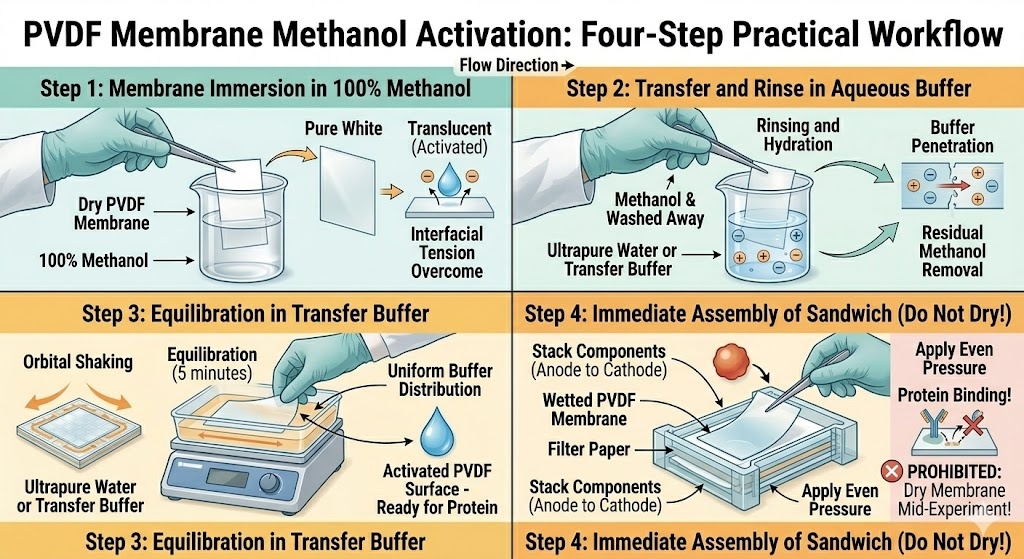
For the transfer buffer itself:
Despite PVDF’s overall superiority in binding capacity and durability, nitrocellulose wins in specific scenarios:
Small proteins (< 25–30 kDa): NC + methanol transfer buffer retains small proteins better than PVDF without methanol. For targets like histones (11–17 kDa), β-actin (42 kDa, near the boundary), and cytokines (8–25 kDa), NC performs comparably or better.
Routine, single-use applications with abundant proteins: If the target is highly expressed, background noise matters more than sensitivity — and NC gives lower background. For a routine quality control blot on a high-expression protein with no reprobing, NC is cheaper and simpler.
No methanol tolerance: Some labs avoid methanol for safety, waste disposal, or because their transfer system is incompatible with high-methanol buffers. NC eliminates this concern entirely.
Nucleic acid detection (Southern/Northern blots): NC is compatible with DNA and RNA hybridization. PVDF is not suitable for nucleic acid blotting.
Dot blotting and slot blotting: NC is the historical standard for these applications and remains widely used.
Mass spectrometry downstream analysis: If you intend to excise protein bands and send them for LC-MS/MS identification or sequencing, PVDF is the only compatible membrane. Nitrocellulose is incompatible with Edman degradation (protein sequencing) and with most MS sample preparation protocols.
Fluorescence-based Western blot: Low-fluorescence PVDF is the only membrane format compatible with NIR fluorescence multiplexing. NC autofluorescence makes it unusable.
High molecular weight proteins (> 100 kDa): Consistent, high-quality bands for large targets (e.g., mTOR at 289 kDa, titin at 3,000+ kDa) require PVDF with a low-methanol or methanol-free transfer buffer.
Multiple reprobing cycles: Any experiment design requiring more than two rounds of stripping and re-detection should use PVDF.
Long-term membrane storage: PVDF membranes can be stored dry at room temperature and rehydrated months or years later with no signal loss. NC degrades with time and storage.
| Problem | Likely Membrane | Cause | Fix |
|---|---|---|---|
| No bands on PVDF | PVDF | Membrane dried during experiment; activation skipped | Re-wet in methanol; never let PVDF dry mid-experiment |
| High background with fluorescence | NC or standard PVDF | Autofluorescence | Switch to low-fluorescence PVDF |
| Weak signal for large protein (> 100 kDa) on NC | NC | Methanol impairs large protein transfer | Switch to PVDF, use low-methanol transfer buffer |
| Signal loss after stripping NC | NC | Mechanical fragility of unsupported NC | Switch to PVDF or supported NC |
| Weak bands after methanol pre-wet of PVDF | PVDF | Buffer not equilibrated after methanol; membrane partially dry | Ensure full 5-min equilibration in transfer buffer |
| Small proteins missing from membrane | Either | Wrong pore size (0.45 µm) | Use 0.2 µm pore size for proteins < 20 kDa |
| Uneven transfer across membrane | Either | Uneven contact with gel; air bubbles | Roll out air bubbles; ensure even pressure in transfer cassette |
Use nitrocellulose if:
Use PVDF if:
When it genuinely does not matter: Both membranes produce comparable results for abundant, medium-MW proteins (30–80 kDa) detected by chemiluminescence with a single antibody. If your target is β-actin, GAPDH, or another highly expressed housekeeping protein at normal loading amounts, either membrane works. Use whatever is already in the lab.



86-15267462807


86-15267462807
